Condition
Knee osteoarthritis
Wear across the knee joint that builds over years. The stage you are at changes which treatments are worth discussing, and moderate is the window where most options are still open.
This page explains the condition. It does not recommend a treatment, because that depends on your joint rather than your diagnosis label.
In plain English
What is actually happening
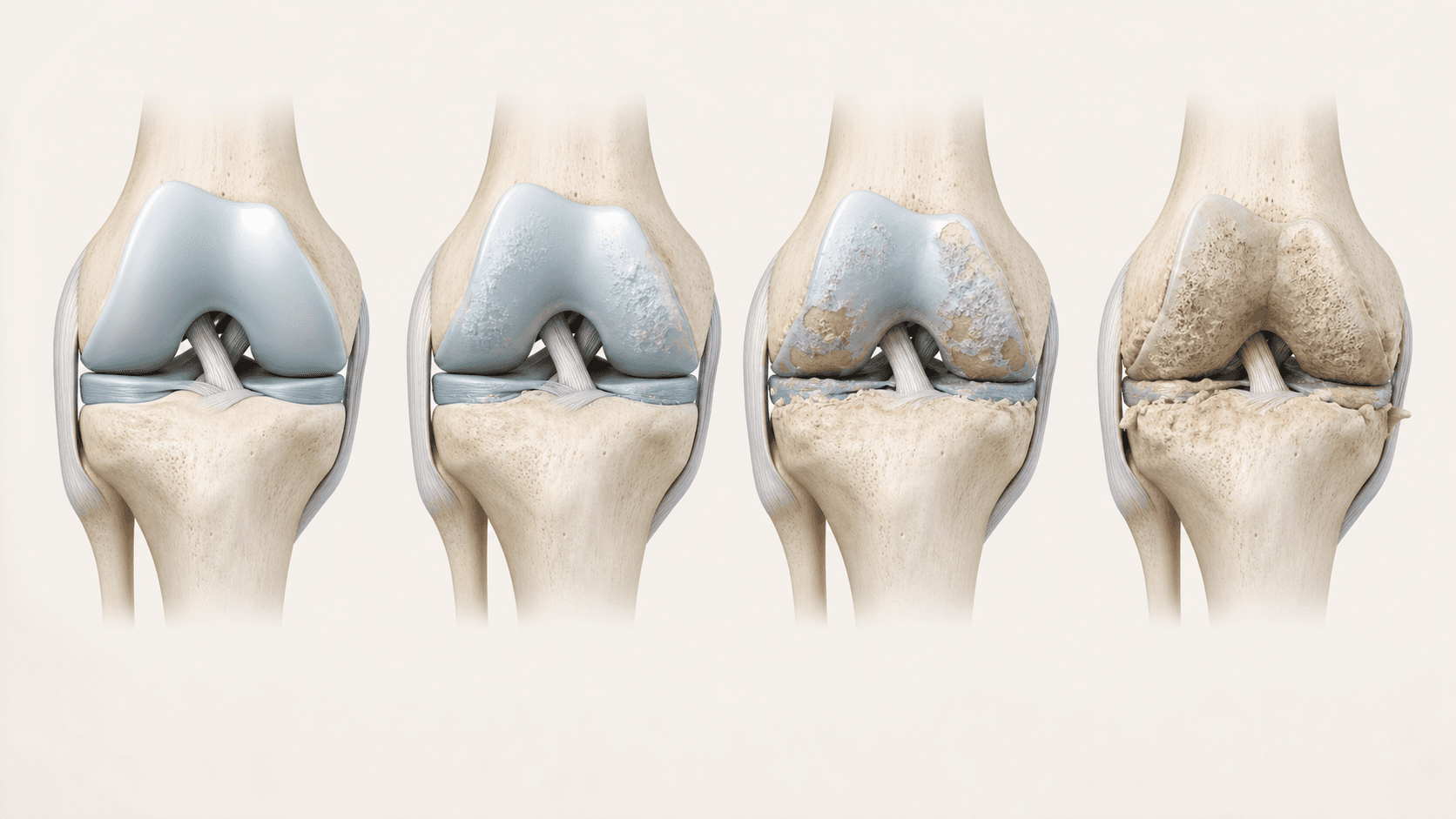
Cartilage is the smooth, glassy surface covering the ends of the bones inside your knee. It lets the joint move almost without friction. In osteoarthritis it gradually thins and roughens.
Cartilage itself has no nerve supply, which is why the wear is not what hurts. The pain comes from what happens around it: the joint lining becomes inflamed, the bone underneath takes more load than it was built for, and the joint space narrows.
That is also why symptoms and scans so often disagree. Two people with near-identical X-rays can have completely different levels of pain, and the treatment decision follows the person more than the picture.
Inside the joint
Where the pain actually comes from
Cartilage has no nerve supply, so the wear itself is painless. Select a marker to see what each part of the joint contributes.
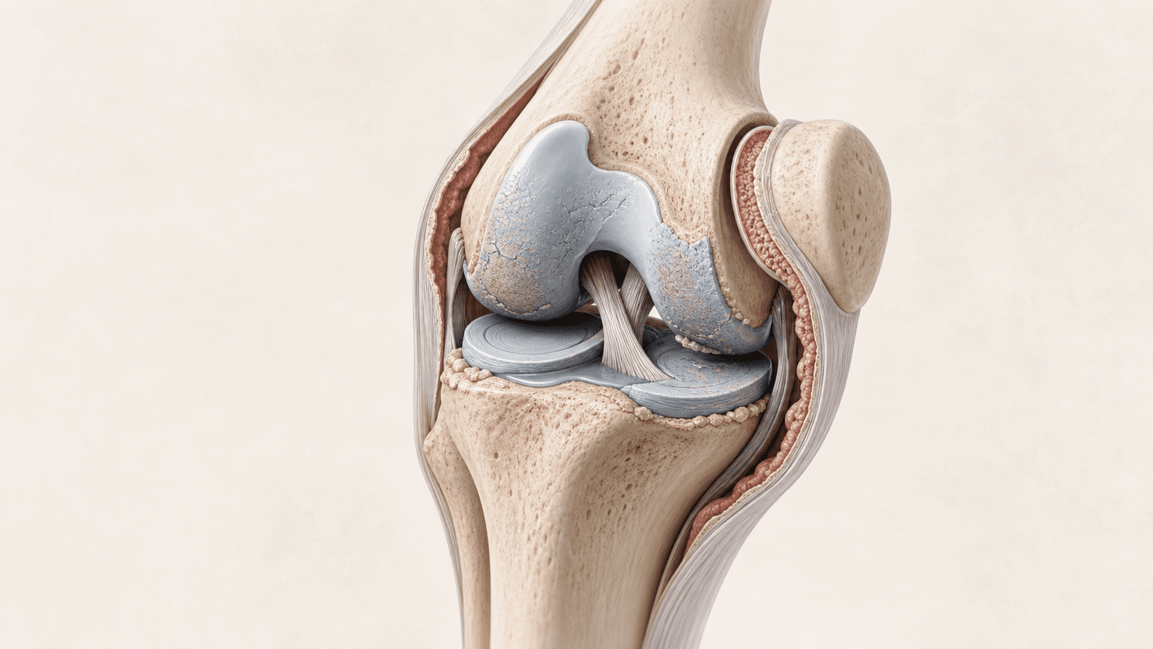
01 · Cartilage thinning
The smooth surface covering the ends of the bones wears down. It has no nerve supply, so the wear itself is painless.
02 · Narrowed joint space
As cartilage thins, the gap between the bones closes. This is the part an X-ray can actually show.
03 · Irritated joint lining
The lining inflames and thickens. This is where a lot of the pain, heat and swelling actually comes from.
04 · Marginal bone change
Small bony growths form at the edges of the joint as it tries to spread load over a wider area.
Common symptoms
What people usually describe
You will not have all of these, and having them does not confirm the diagnosis.
Pain on load
Worse going up or down stairs, on slopes, or after being on your feet for a while.
Morning stiffness
Stiff for the first half hour or so, then easing as you get going.
Swelling and heat
The joint puffs up or feels warm, particularly after activity. That is the lining, not the cartilage.
Grinding or creaking
A grating sensation through the joint as the surfaces lose their smoothness.
Losing distance
Walking less than you used to, or planning around the knee rather than forgetting about it.
Disturbed sleep
Pain at night or being woken by the joint is a meaningful marker of severity.
Or is it something else
How this differs from similar problems
Locking or giving way
A knee that genuinely catches, locks or lets you down points at something mechanical, such as a meniscal tear or a loose fragment. Injections do not fix mechanical problems.
Front-of-knee pain only
Pain focused behind the kneecap, worse on stairs and after sitting, may be patellofemoral osteoarthritis, which is a distinct diagnosis with different mechanical drivers.
Rapid onset with heat
A joint that becomes hot, red and severely painful over hours rather than years needs urgent medical assessment, not an injection clinic.
Imaging
Why the scan matters, and where it misleads
An X-ray shows bone. Cartilage is invisible on plain film, so an X-ray infers wear from how narrow the joint space has become. At moderate osteoarthritis this substantially understates the actual cartilage damage.
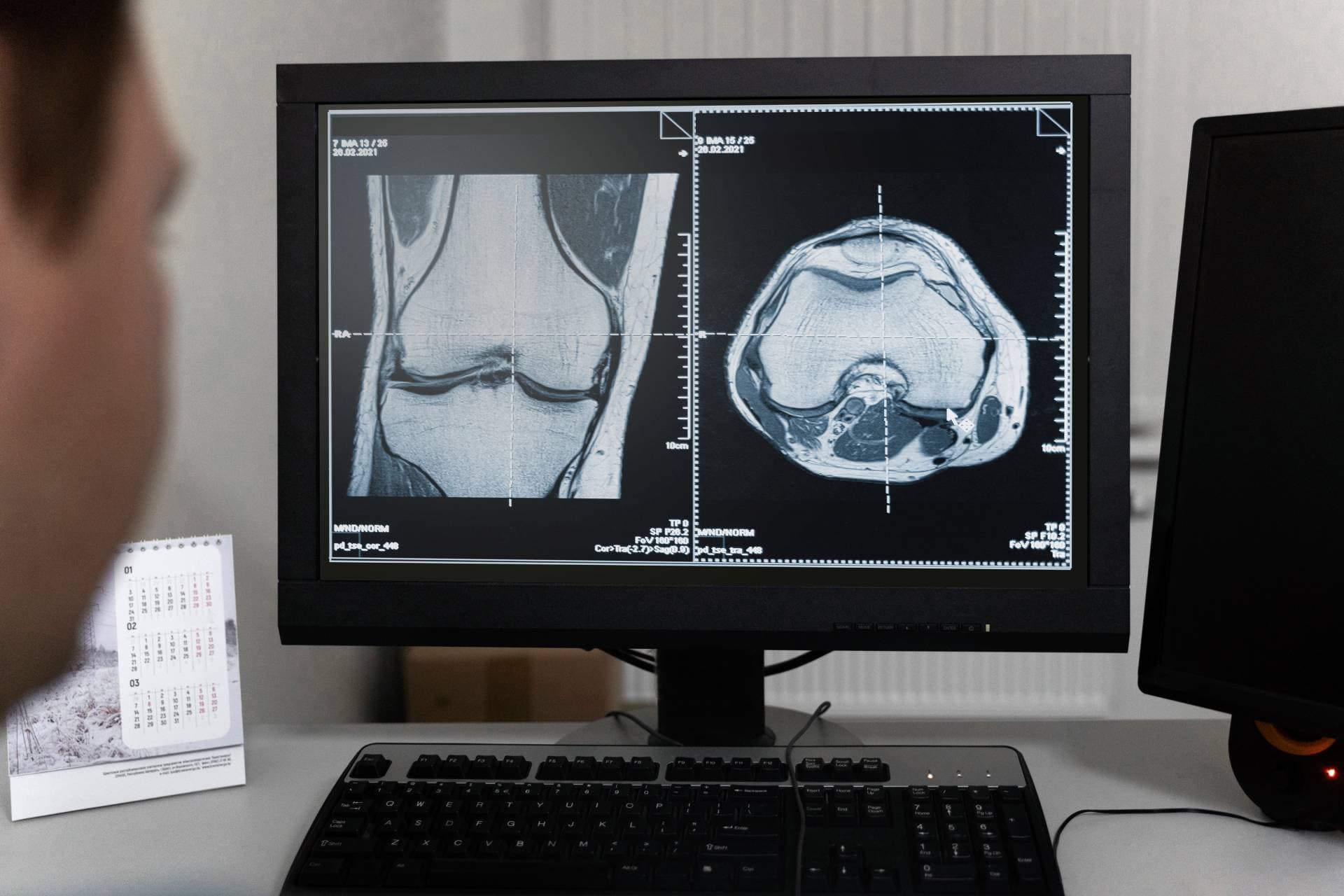
An MRI shows the cartilage itself, the joint lining and the bone underneath it. It also picks up bone marrow lesions, which are areas of inflammation in the bone beneath the cartilage and a known cause of pain that is disproportionate to the damage visible elsewhere.
This is why we ask what imaging you already have before recommending anything. It is also why a scan more than a year old often needs repeating: the decision is built on what the joint looks like now.

Where it sits
On the spectrum of wear
Moderate is the critical window. Waiting for symptoms to worsen closes options rather than preserving them.
What may be discussed
Treatments that tend to come up
Not a recommendation. These are the options a doctor may raise for this condition, and which one fits is decided on examination and imaging.
Lubricates
Hyaluronic acid
The usual first injection: lubricates the joint, lowest cost, around six months of relief.
Cushions
Arthrosamid®
Cushions the joint and settles the inflamed lining, aiming at years from one injection.
Rebuilds
ChondroFiller Injection®
Where the goal is to support the worn surface rather than manage the symptoms around it.
Possibly none of them
An assessment first
Where the pattern does not fit an injection at all, which is a legitimate outcome.
Book an assessmentLimits
When an injection is not enough
We offer four injections and nothing else. These are the situations where the honest answer sits outside them.
- The joint locks, catches or gives way, which points at a mechanical problem
- The knee is severely worn and the aim is to fix it rather than manage it
- Pain at rest and at night that no longer responds to anything
- Significant deformity or instability, which changes how the joint is loaded
Urgent
When to seek help sooner
These are not injection-clinic problems. If any apply, contact your GP, NHS 111 or an emergency department rather than booking with us.
- A hot, red, severely painful joint developing over hours: seek urgent medical assessment
- Fever alongside a swollen joint
- A joint that gives way and causes falls
- Sudden inability to straighten or bend the knee
- Unexplained weight loss or night pain alongside joint symptoms
Questions
Common questions
- Does osteoarthritis always get worse?
- Not at a fixed rate, and not for everyone. It varies enormously. What is true is that acting while there is still cartilage to work with keeps more options open than waiting does.
- My X-ray was not that bad but I am in a lot of pain.
- That is common and it is not in your head. X-rays understate cartilage damage, and much of the pain comes from the joint lining and the bone beneath the cartilage rather than from the cartilage itself.
- Should I try physiotherapy first?
- Usually yes. An injection is generally appropriate after exercise, weight management, physiotherapy and pain relief have been genuinely tried and found inadequate, rather than because a scan shows osteoarthritis.
- I have been told I need a knee replacement. Is it too late for an injection?
- Not necessarily. Being told you may need a replacement is not the same as being told you need one now, and it is worth understanding what an injection can and cannot offer at that stage before deciding.
- I am under 60. Does that change anything?
- It can. Knee implants last around 15 to 20 years, so a replacement before 60 makes revision surgery likely while you are still relatively young. That is a real argument for exploring what else is available first.
Related
Find out what your joint actually needs.
An assessment looks at the joint, your imaging and what you want back, then tells you which of the four fits, or whether none of them does.