Condition
Diffuse cartilage wear
Thinning spread across the whole joint surface rather than sitting in one spot. Harder to treat than a contained defect, but not the end of the conversation.
This page explains the condition. It does not recommend a treatment, because that depends on your joint rather than your diagnosis label.
In plain English
What is actually happening
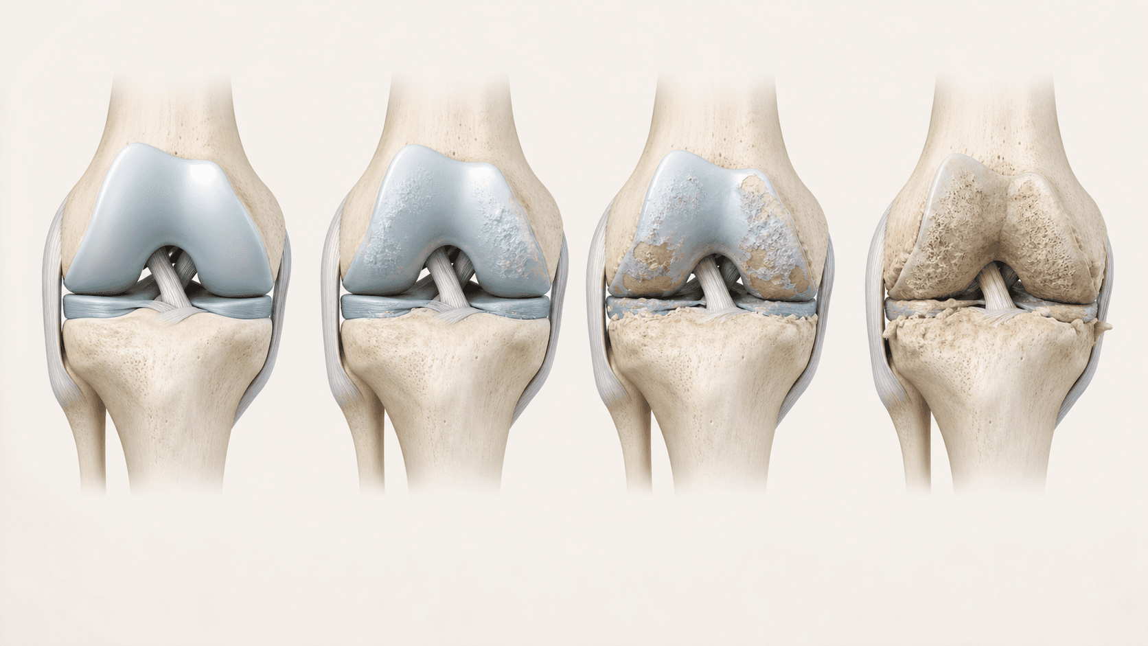
Rather than one hole in the surface, the cartilage has thinned broadly across the joint. There is no single target to aim at, which is precisely what makes it harder to treat.
This is the pattern most people mean when they say osteoarthritis. The joint space narrows, the lining becomes irritated, and the bone underneath starts carrying load it was not designed for.
It matters that this is a spectrum rather than a switch. There is a real difference between a joint with thinned but continuous cartilage and one that has genuinely run out. Both get called worn; they are not the same problem.
Inside the joint
Where the pain actually comes from
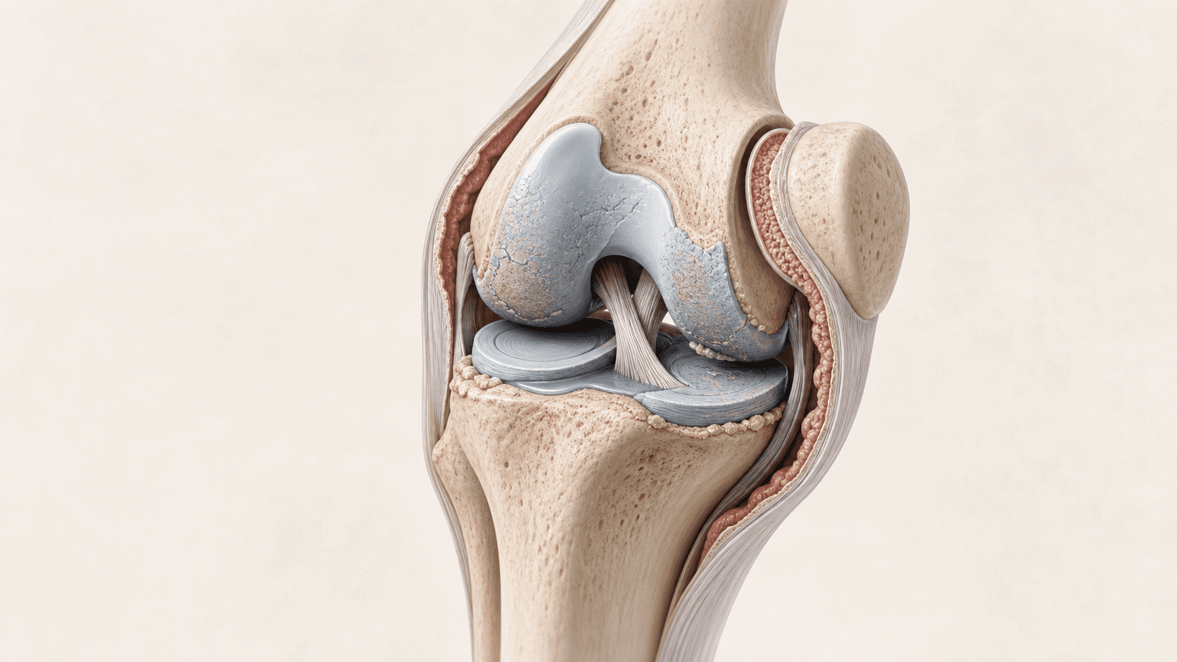
Cartilage has no nerve supply, so the wear itself is painless. Select a marker to see what each part of the joint contributes.
01 · Cartilage thinning
The smooth surface covering the ends of the bones wears down. It has no nerve supply, so the wear itself is painless.
02 · Narrowed joint space
As cartilage thins, the gap between the bones closes. This is the part an X-ray can actually show.
03 · Irritated joint lining
The lining inflames and thickens. This is where a lot of the pain, heat and swelling actually comes from.
04 · Marginal bone change
Small bony growths form at the edges of the joint as it tries to spread load over a wider area.
Common symptoms
What people usually describe
You will not have all of these, and having them does not confirm the diagnosis.
Widespread ache
Pain through the joint generally, rather than in one identifiable spot.
Stiffness after rest
Stiff getting going in the morning or after sitting, easing as you move.
Grinding through range
A gritty or creaking sensation as the joint moves, not just at one angle.
Reducing distance
Walking less than you did, and planning routes around the joint.
Recurrent swelling
The joint puffs up regularly rather than only after unusual activity.
Night pain
Being woken by the joint is a meaningful marker that things have progressed.
Or is it something else
How this differs from similar problems
A single painful spot
Pain you can point to precisely, in an otherwise good joint, is more likely a focal defect, which has a better outlook.
Mechanical locking
True locking suggests a fragment or a tear on top of the wear, and that changes the plan.
Inflammatory arthritis
Multiple joints involved, prolonged morning stiffness or systemic symptoms point somewhere else entirely and need a different specialist.
Imaging
What imaging tells you here
An X-ray shows how far the joint space has closed, which is genuinely useful at this end of the spectrum. It is the measure that separates worn from bone on bone.
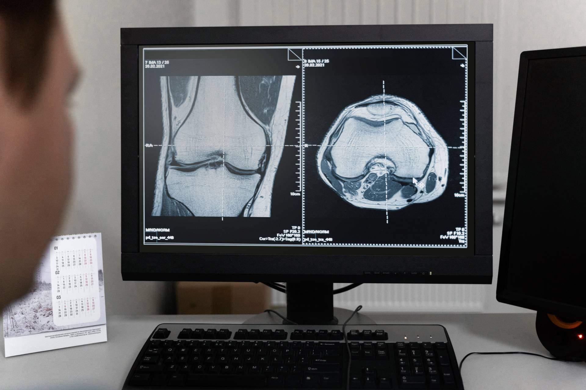
An MRI adds what the X-ray cannot: how much cartilage is actually left, whether the lining is inflamed, and whether there are bone marrow lesions driving pain out of proportion to the visible damage.
At this stage imaging is not just about deciding which injection. It is about the honest question of whether an injection is the right move at all, or whether a joint replacement deserves to be on the table.

Where it sits
On the spectrum of wear
Options narrow as this progresses, which is the argument for having the conversation sooner rather than later.
What may be discussed
Treatments that tend to come up
Not a recommendation. These are the options a doctor may raise for this condition, and which one fits is decided on examination and imaging.
Cushions
Arthrosamid®
Cushions the joint from the inside and calms the lining. Aims at years of relief from one injection.
Rebuilds
ChondroFiller Injection®
Lays a scaffold across broad surface wear rather than filling a single hole. The widest range of the four.
Lubricates
Hyaluronic acid
Lower cost and lower commitment, though it gives less in a more worn joint.
Possibly none of them
An assessment first
Where a joint replacement is genuinely the better answer, you should be told that plainly.
Book an assessmentLimits
When an injection is not enough
We offer four injections and nothing else. These are the situations where the honest answer sits outside them.
- The joint has effectively run out of cartilage and is bone on bone
- Pain at rest and at night that nothing touches any more
- Deformity or instability that changes how the joint carries load
- Function has deteriorated to the point where a replacement would restore more than an injection could
Urgent
When to seek help sooner
These are not injection-clinic problems. If any apply, contact your GP, NHS 111 or an emergency department rather than booking with us.
- A hot, red, severely painful joint over hours, which needs urgent assessment
- Fever with a swollen joint
- Giving way that causes falls
- Unexplained weight loss or night pain alongside joint symptoms
Questions
Common questions
- Is bone on bone the same as diffuse wear?
- No, and the distinction is worth insisting on. Diffuse wear means thinned cartilage across the surface. Bone on bone means it has largely gone. Both get called worn, and they have different options.
- I have been told nothing can be done. Is that right?
- Sometimes an injection genuinely is not the answer and we will say so. But being told a joint is worn is not the same as being told nothing is available, and it is worth understanding the difference.
- Can I delay a replacement?
- Injections can delay a replacement, with published estimates ranging from a few months for hyaluronic acid to a few years for a hydrogel. How long depends on the joint, not on the injection alone.
- Will an injection make a future replacement harder?
- Having had an injection does not rule out a replacement later. This is a reasonable question to put directly to the doctor assessing you.
Related
Find out what your joint actually needs.
An assessment looks at the joint, your imaging and what you want back, then tells you which of the four fits, or whether none of them does.