Condition
Focal cartilage defects
One contained area of damage in an otherwise sound joint. The most treatable pattern there is, and the one where acting early makes the most difference.
This page explains the condition. It does not recommend a treatment, because that depends on your joint rather than your diagnosis label.
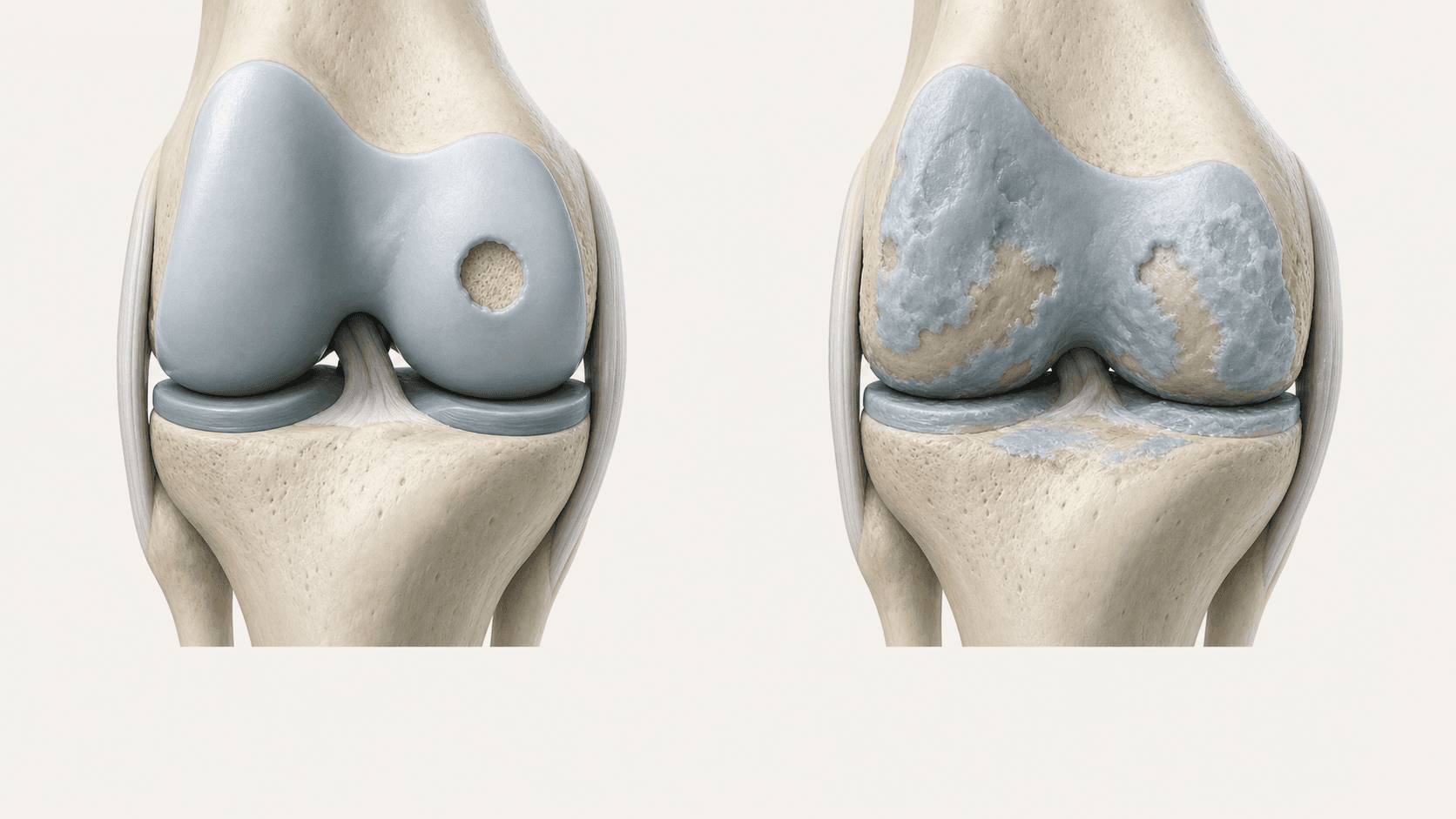
In plain English
What is actually happening
A focal defect is a discrete area where the cartilage surface has been lost, with reasonably healthy cartilage around it. Think of a pothole in an otherwise sound road rather than a road that has worn out everywhere.
They are commonly the result of an injury: a twist, an impact, or a fragment that has come away. They can be silent for a long time and then start causing trouble when the joint is loaded harder.
The reason this pattern matters is that the joint around the defect is still doing its job. There is intact surface to build against, and the mechanics are usually sound. That makes it the most favourable pattern for a treatment that works on the surface.
Common symptoms
What people usually describe
You will not have all of these, and having them does not confirm the diagnosis.
Pain in one place
You can often point to where it hurts, rather than describing a general ache.
Worse on load
Deep flexion, stairs, squatting and impact provoke it more than flat walking.
Intermittent swelling
The joint swells after harder use and settles with rest.
Catching
A sense of something snagging as the joint passes through a particular angle.
A specific incident
Many people can name the twist or impact that started it.
Otherwise a good joint
No general stiffness, no grinding through the whole range: just this one spot.
Or is it something else
How this differs from similar problems
Wear across the whole surface
If the joint is stiff and grinding throughout its range, the picture is diffuse wear rather than a focal defect, and a different approach applies.
A meniscal tear
Tears also catch and swell, but they sit in the cushion between the bones rather than in the joint surface. Imaging distinguishes them.
Pain from instability
If the joint gives way, the ligament problem needs addressing first. Resurfacing a joint that is not stable does not last.
Imaging
Why an MRI is essential here
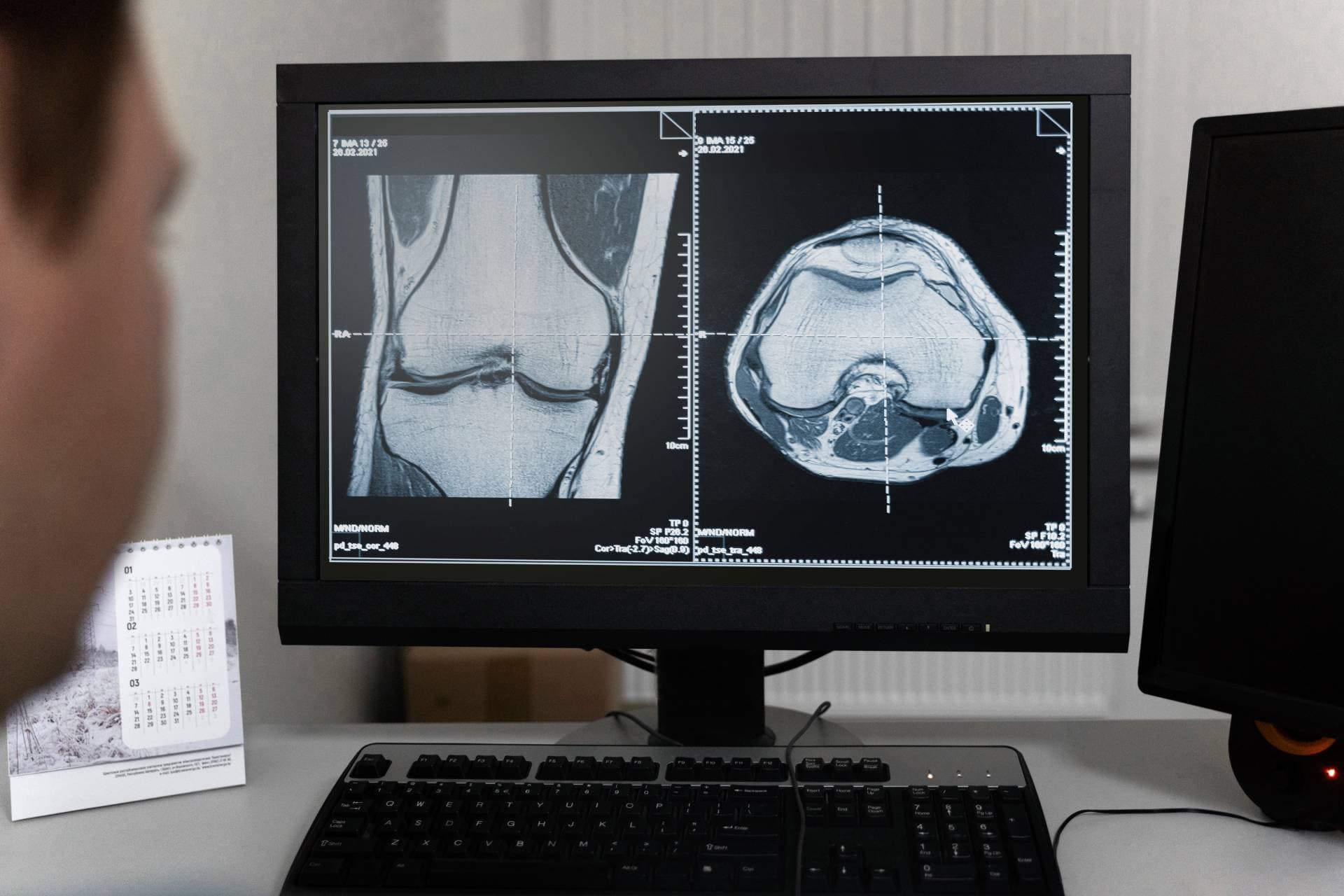
For focal defects, imaging is not a formality. The treatment plan is built directly on where the defect is, how deep it goes, and how big it is.
An MRI also confirms the thing that makes this pattern favourable: that the rest of the joint is intact and mechanically sound. If it is not, the plan changes.
It shows whether the bone underneath is involved, which affects both the approach and the realistic expectation of what a treatment can achieve.

Where it sits
On the spectrum of wear
A focal defect sits at the treatable end. Left long enough, the surrounding surface starts to suffer too.
What may be discussed
Treatments that tend to come up
Not a recommendation. These are the options a doctor may raise for this condition, and which one fits is decided on examination and imaging.
Rebuilds
ChondroFiller Injection®
The scaffold approach was originally designed for exactly this: a contained defect for your own cells to rebuild into.
Regenerates
Mytocel MSK
Your own cartilage cells, where the defect is early and the surrounding joint is healthy.
Lubricates
Hyaluronic acid
Symptomatic help while a decision is made, though it does nothing to the defect itself.
Possibly none of them
An assessment first
Where mechanical problems sit alongside the defect and need dealing with first.
Book an assessmentLimits
When an injection is not enough
We offer four injections and nothing else. These are the situations where the honest answer sits outside them.
- A loose fragment is moving inside the joint and causing true locking
- The joint is unstable, so any resurfacing would be working against the mechanics
- Significant malalignment concentrates load directly onto the defect
- The bone beneath the defect is substantially involved
Urgent
When to seek help sooner
These are not injection-clinic problems. If any apply, contact your GP, NHS 111 or an emergency department rather than booking with us.
- A joint locked in position that will not straighten
- Sudden severe pain after an injury with inability to weight-bear
- A hot, swollen joint with fever, which needs urgent assessment
- Repeated giving way causing falls
Questions
Common questions
- Is my defect too big to treat?
- Size is less decisive than people assume. What matters more is whether the joint environment can support a repair response. That is judged on imaging and examination, not on a measurement alone.
- Do I need surgery for this?
- Not necessarily. For focal, stable defects in a mechanically sound joint, an outpatient ultrasound-guided scaffold injection can do the job without surgical repair.
- Am I too old?
- There is no upper age limit on the injection itself. Suitability is about the joint, not the birthday.
- What happens if I leave it?
- A defect does not heal on its own. Over time the edges and the surrounding surface come under more strain, and a contained problem can become a diffuse one, which is harder to treat well.
Related
Find out what your joint actually needs.
An assessment looks at the joint, your imaging and what you want back, then tells you which of the four fits, or whether none of them does.